Tuesday, December 6, 2011

2.10 Effect of pH on the rate of reaction

 Understand how the functioning of enzymes can be affected by changes in pH

1. Lower PH (acidic) = the rate of reaction gradually decreases. 
Optimum PH = maximum rate = rate of reaction decreases. 
Higher Ph (alkaline conditions) = rate of reaction gradually decreses. 

Graph is more symmetrical. 

Acidic and Alkaline conditions change the shape of the ACTIVE SITE and this slows the reaction. 

2.9 Effect of Temperature on the rate of reaction

 Understand how the functioning of enzymes can be affected by changes in temperature

1. a) Increase in temp = increase in average KE of particles. 
b) 
increase in average KE of particles = more frequent collisions = more fruitful reactions. 
2. Particles = Substrate and the Enzyme 
 Reaction = E+S ---> ES(complex)--> E + Product formed. 

Low temp = low KE, few complexes formed and product will be formed slowly (slow rate of reaction)
Higher temp = KE of S and E increase --> more complexes formed = more product formed. 
Optimum temp (peak of curve) = rate of reaction declined very quickly because KE changes the shape of the ACTIVE SITE of the enzyme = denatured. (cannot produce any more product).

2.8b Enzyme Reactions

Understand the role of enzymes as biological catalysts in metabolic reactions. 

1. (Oxidation of oxygen) Respiration C6 H12 O6 + O2 ---> Energy + CO2 + H2O
    Substrate                                          Released          Product

2. Combustion (adding energy) = Without an enzyme - to break down glucose ---> carbon dioxide and water - we have to put energy into the glucose molecule bonds before they break and release a lot of energy.

3. Energy of Activation = initial energy added to glucose molecules.

The shape of the active site is affected by pH. This is why enzymes will only work at a specific pH, as well as a specific temperature. Change the pH (acidic) and the enzyme stops working.
Increasing the temperature to 60°C will cause a permanent change to the shape of the active site. This is why enzymes stop working when they are heated. We say they have become denatured.

Biological systems overcome the energy of activation by THE ENZYME.

Wednesday, November 30, 2011

2.8a Enzymes

2.8 understand the role of enzymes as biological catalysts in metabolic reactions

28a Enzymes

1. Catalyst = makes the reaction occur faster under moderate conditions. 
2. Metabolical reactions = biological reactions taking place in a cell, building molecules up or breaking them down. 
3. Lock and Key Hypothesis 
Enzymes have a particular shape 
Enzymes are 
proteins

a)  Enzyme

b) Active site - where the substrate (c) fits in. Called: COMPLEMENTARY 
(d) Activated Complex (substrate in active site)
(e) Products emerge from the enzyme (substrate has been broken down into two products) - (f)

The  
Enzyme remains unchanged

Substrate -----> Product 

2.7 Lab Tests for glucose and starch

2.7 describe the tests for glucose and starch

1. Glucose (C6 H12 O6) positive test 
a) Take glucose powder and dissolve it in a test tube 
b) Add  Benedict's Reagent (blue) 
c) Take test tube and place in a water bath to boil (60-70C) for 2-3 minutes
d) Remove test tube
Result = colour change from blue to orange.


2. Starch positive test 
a) In solid form = white powder
b) Place some white powder into a spotting tile 
c) Add Iodine Solution (brown)
Result = colour change from brown to dark blue/black colour

2.6 Biological Molecules Structure

2.6 describe the structure of carbohydrates, proteins and lipids as large molecules made up from smaller basic units: starch and glycogen from simple sugar; protein from amino acids; lipid from fatty acids and glycerol

1. Carbohydrates (CHO) ---> Sugars (glucose) combine to form STARCH (plants) and GLYCOGEN (animals)


2. Proteins (CHON) ---> Amino Acids join together in long chains which join together to make proteins.
Broken down they form smaller molecules (the amino acids)


3. Lipids (CHO) ---> (have 2 different kind of molecules) --> Glycerol + Fatty acids which join together to form Lipids.

2.5 Elements in biological molecules

2.5 recall the chemical elements present in carbohydrates, proteins and lipids (fats and oils)

1. Carbohydrates --> Carbon, Hydrogen, and Oxygen ---> Sugars + Polysaccharides (starch)
2. Proteins           ---> Carbon, Hydrogen, Oxygen, and Nitrogen ---> Amino Acids + Proteins
3. Lipids              ----> 
Carbon, Hydrogen, and Oxygen ----> Fats (animals) and Oil (plants) 

Tuesday, November 29, 2011

2.4 Differences between Plant and Animal cell

2.4 describe the differences between plant and animal cells.

Animal Cell: 
Cell membrane
Nucleus
Cytoplasm 

Shape: Rounded
Store: Glycogen molecule (carbohydrate)


Plant Cell:
Cell MembraneNucleusCytoplasm
Cell wall
Chloroplasts
Shape: regular shape
Central Vacuole
Store: Starch molecule (carbohydrate)

Functions of parts of a call

1. Plant Cell 
Vacuole = Stores molecules (amino acids, sugars etc)
Chloroplasts = Where photosynthesis occurs
Cell wall = Provides the protection and support for the cell. 
2. Animal cell

Cell Membrane = controls what goes and leaves the cell
Nucleus = Controls what the cell does
Cytoplasm = where chemical reactions take place eg: respiration.

Sunday, November 27, 2011

2.2 Cell structure

Recognize cell structures, including the nucleus, cytoplasm, cell membrane, cell wall chloroplast and vacuole.  
N = nucleus
CM = Cell membrane
C= Cytoplasm
V= Vacuole
CW= Cell wall

Sunday, November 20, 2011

2.1 Levels of Organisation

2.1 describe the levels of organisation within organisms: organelles, cells, tissues, organs and systems.
example suing Plant.
1. System: Composed of a group of organs  working together on eg: Plant cell; Reproduction, Photosynthesis, Transport.

2.  Organ:  Group of different Tissues working together.
Leaf: 3 different Functions: Photosynthesis, Gas Exchange and Transpiration.

3. Tissue - Group of cells of the same kind = they have the same shape and function.
eg: Phloem - Cell that transports "sap"
eg: Xylem - Transports water and minerals.
eg: Plaside - Carries out P/S


4. Cells: multicelluler organinisum.
a) In development of the organism, the cell specializes: they switch on genes which change their shape giving the cell a particular function: This is called  Cells differentiation 

Monday, November 7, 2011

2.75 Urine

Recall that urine contains water, urea and salts 

1. Brain influences the the functioning of the kidneys
2. Kidneys filter out blood
3. Molecules removed come out the ureter and collect in the bladder as urine
4. Urine contains salt, water and urea --- which affects the composition of the tissue fluid = Osmoregulation
5. Removal of urea = excretion of metabolic waste.
- composition of salt, water and urea varies and depend upon conditions in which a person is operating.

Sunday, November 6, 2011

2.74 ADH.

Describe the role of ADH in regulating the water content of the blood. 

1. ADH (anti - directic hormone) is produced in the brain - located in the Hypothalamus region of the brain. ADH flows through the blood stream and targets the kidneys.


2. Function of ADH: to control and alter the quantity/composition in the blood. - Making it more or less concentrated.

3. ADH targets --> collecting duct


Affect: allows more water to come out of the collecting duct. (collecting duct = where water is selectivly reabsorbed into the blood) - ADH increases the quantity of the water being removed.

4. ADH secretion = more water goes back into the blood, urine becomes more concentrated and the volume decreases. 

2.73 Glucose reabsorption


Understand that selective reabsorption of glucose occurs at the proximal convoluted tubule. 

1. Selective reabsorption = molecule is selected --> (glucose)
    reabsorption - glucose will go from the Glomeral filtrate and then go back into the blood

2. Filtration occurs in the Bowmans Capsule - The filtrate containing glucose, water, salts and urea (formed from the high pressure of blood) - Water is removed back into the blood in the collecting duct

3. Urine coming out of the Nephron - does not contain glucose. (if glucose is present in urine this is an indication of a condition called Diabetise).

4. Glucose is in the fluid in the first Proximal (1st) Convoluted (twisted) tubule - glucose is removed here and taken back into the blood.

5. Glucose is selectively reabsorbed into the blood in the Proximal Convoluted Tubule (no glucose in Urine)


2.72 Water re-absorption.

Understand that water is reabsorbed into the blood from the collecting duct. 

1. Bowman's capsule: where ultra filtration occurs.

a)Blood goes into the kidney at high pressure
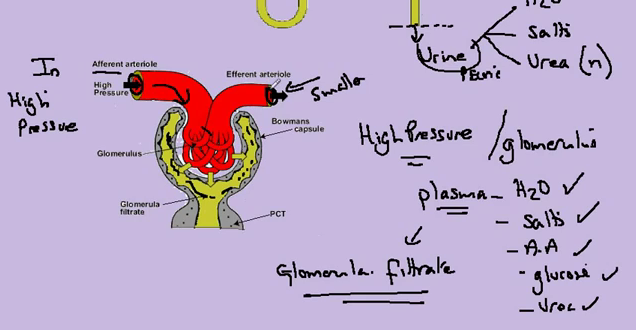
b)The dissolved contents of the blood are forced into the Bowman's capsule (Glomerula filtrate) --> glucose, water, salt, and urea. 
c) When filtration occurs, too much water is filtrated. The filtrate passes along the tubule and when it reaches the collecting duct and passes through - water is removed from the filtrate as it goes down the medula. The water is then returned back into the blood vessels.
d) Water has been selected - and reabsorbed into the blood.

2.71 Ultrafiltration


Describe ultrafiltration in the Bowman's capsule and the composition of the glomerular filtrate. 

1. Nephron - carries out the filtration of blood --> filtered blood (clean)
                                                                            --> Urine (waste)
2. Urine is composed of: Water, Salts, and Urea (nitrogen waste - toxic) 

3. Bowman's Capsule: where ultra-filtration begins.
a) Blood arrives in the kidney in the Afferent arteriole - at high pressure - (with a wide blood vessel).
b) Blood passes through the Glomerulus and pases out the Efferent arteriole - (with a narrow blood vessel.)
c) Blood pressure increases in the Glomerulus.
d) High pressure forces the plasma (liquid in blood - water, salts, amino acids, glucose and urea) out of the blood vessel into the inside of the Bowman's Capsule - This is called "Glomerula Filtrate."

2.70 Nephron Structure

Describe the structure of a Nephron to include Bowman's Capsule and glomerulus, convoluted tubules, loop of Henlé and collecting duct. 


  1. Renal Vein
.Carries blood away from the kidney and back to the right hand side of the heart. Blood in the kidney has had all its urea removed. (Urea is produced by your liver to get rid of excess amino-acids).
Blood in the renal vein also has exactly the right amount of water and salts. This is because the kidney gets rid of excess water and salts. The kidney is controlled by the brain. A hormone in our blood called  ADH  is used to control exactly how much water is excreted.

  1. Renal Artery
This blood vessel supplies blood to the kidney from the left hand side of the heart. This blood must contain glucose and oxygen. Blood in the renal artery must have pressure to be able to filter the blood.
Blood supplied to the kidney contains a toxic product called urea which must be removed from the blood. It may have too much salt and too much water. (The kidney removes these excess materials)

  1. Pelvis
This is the region of the kidney where urine collects. 

  1. Ureter
The ureter carries the urine down to the bladder and is stored there until excreted.
 E.     Medulla
The medulla is the inside part of the kidney (green) (real life: dark red) This is where the amount of salt and water in your urine is controlled. It consists of billions of loops of Henlé. ADH makes the loops work harder to pump more sodium ions. The result of this is that very concentrated urine is produced.


  1. Cortex
The cortex is the outer part of the kidney. This is where blood is filtered. We call this process "ultra-filtration"  because it only works if the blood entering the kidney in the renal artery is at high pressure.
A glomerulus is a tiny ball of capillaries (found in the cortex). Each glomerulus is surrounded by a "Bowman's Capsule". Glomeruli leak. Things like red blood cells, white blood cells, and platelets  stay in the blood vessels. Most of the plasma leaks out into the Bowman's capsules. 
Most of this liquid, which we call "ultra-filtrate" is re-absorbed in the medulla and put back into the blood.


Thursday, October 27, 2011

2.69 Urinary system

Describe the structure of the urinary system, including the kidneys, ureters, bladder and urethra

1. Each kidney has its own blood supply and tube called the ureter (which leads to the bladder)

2. Ureter carries urine from kidney to the bladder

3. Urine is excreted through the urethra of the penis/vagina.
 

2.68b Osmoregulation

Understand how the kidney carries out its roles of excretion and of osmoregulation 

1. (process) osmoregulation
Osmo = Osmosis
Regulation = to control

2. The tissue fluid which surrounds these cells must be ISOTONIC with the cytoplams of these cells (= the amount of water going into + out must be equal and the cells remain theier function + shape)

3. Blood circulating into the tissueHypertonic (conc) - add too much water to the cells
or
 Hypnotic (dilute) - remove too much water from the cells

4. ISOTONIC conditions is achieved by controlling the composition of blood
- blood forms the tissue fluid
- kidney controls the composition of blood
- excess water + salts are removed

2.68a Excretion

Understand how the kidney carries out its roles of excretion and of osmoregulation 

1. Excretion of Urea -> nitrogen (toxic) excess amino acids

- Blood circulates to the liver and the amino acids are broken down into the molecules --> Urea.
-Urea then circulates to the kidneys
- The kidneys filter the Urea from the blood 
- Urea is added to water --> Urine
- Urine is then collected in the bladder
-Urea is now in the bladder and removed from the body

2.67b Human organs of Excretion

Recall that the lungs, kidneys and skin are organs of excretion 
1. Lungs excrete carbon dioxide
2. Kidneys excrete excess water + urea (nitrogen wastes from amino acides) + salts
3. Skin excrete water + salts (sweat)

OBJ - 2.67a Excretion in plants

Recall the origan of carbon dioxide and oxygen as waste products of metabolism and their loss from the stomata of a leaf 

1. Example of Excretion (in leaf)
Carbon dioxide + Water --> Glucose + Oxygen (Waste molecule)
                                             Release of metabolic waste ^^

2. (Aerobic) Respiration
Glucose + Oxygen --> ATP + Carbon Dioxide + Water
                                                 Release of metabolic waste ^^

Friday, October 7, 2011

3.34 Causes of mutation

Understand that the incidence of mutations can be increased by exposure to ionizing radiation (for example gamma rays, X-rays and ultraviolet rays) and some chemical mutagens (for example chemicals in tobacco).

1. Mutation is the change in the base sequence of the gene.   New alleles are produced
2. what causes mutation?
examples are: a) radiation: X-rays, UV-B rays (sunshine) can cause skin cancer
                      b) chemicals such as tar - they are called mutagenes and cause cancer - carcinogens

3.33 Antibiotic resistance

Understand how resistance to antibiotics can increase in bacterial populations. 


1. Staphylococcus aureus ---> causes skin infections + lung infections

2. Infections can be treated with methecilline (antibiotic) it kills the Staphylococcus aureus. This is the susceptible form. MSSA.

3. Staphylococcus aureus infection increase is  caused by random mutation and when the methecilline (antibiotic) is applied - the bacteria does not die - this is called the resistant form. MRSA
The mutation has created genes in Staphylococcus aureus  that makes it resistant so it doesn't die from the methecilline (antibiotic).

4. Commonly found in hospitals - from treatment of people with wounds. Resulting in the methecilline (antibiotic) no longer working. 

3.32 Types of mutation

Understand that many mutations are harmful but some are neutral and a few are beneficial. 


1. Impact of mutation on new alleles. 

Process of mutation -->


Beneficial effect - eg: improve efficiency of an enzyme
Neutral effect - eg: ( no particular thing) but with time due to environmental change - can effect it.
Harmful effect - eg: mutation produces production of n enzyme that doesn't work.

Thursday, October 6, 2011

Obj - 3.31 Evolution

Describe the process of evolution by means of natural selection 


1. Evolution : a) change in the form of organisms - b) change in frequency of alleles. 
2. Natural slelection: is the system of evolution. (introduced by Charles Darwin)
3. Change in allele frequency by natural selection:
example: Staphylococcus aueurs.
*Susceptible: likely to be harmed by 
 2 features of classic process of  natural selection 
1. Random mutation - produces the MRSA resistant form
2. Non-random selection - due to the antibiotic which is selecting the MRSA (methecilline antibiotic) survive and the MSSA to be selected and killed.

3.30 Mutation

Recall that mutation is a rare, random change in genetic material that can be inherited. 
*Allele = form of gene

A change occurs which produces a new version of an allele which produces a different portion.
And resulting a different effect on the phenotype.

Different allele's exist due to mutation
Mutation - changes the base sequence of the gene.

3.29 Species Variation

Understand that variation within a species can be genetic, environmental or a combination of both.

1. variation = difference we can see in the phenotype (how things appear)
it is possible to count and measure these differences in graphic form.

An individual has a phenotype --> genotype --> which is modified by the environment.

variation pop = variation genotype = variation environment

Differences in the appearance of individuals in a species population  is because those individual have different genotypes and are living+surviving in different environments.

Graph "1" - suggests variation in the pop (species) is due to the variation in genotypes. (environment is normal here)
Discontinuous variation --->


Graph "2" suggests variation in the pop and variation in species.
The variation here is caused by genetic variation in the genotype

eg: Height in humans (affected by your diet)
Combo of genes + the environment.


these groups are modified by the environment which results in a smooth curve.


3rd case: Variation in a population (phenotypic variation) is due to environmental variation. The genes have no role to play in differences in pop.

eg: the home language you speak.

Sunday, September 25, 2011

Definitions

Phenotype: what they look like.
Genotype: the genetic make up of a single cell
Homozygous: same alleles
Heterozygous: a cell having two different alleles of a particular gene(s)

Tuesday, September 20, 2011